DEFINITION OF RWMA
Regional wall motion abnormalities is shortly called as RWMA. It means a part of left side chamber of the heart (usually left ventricle) is not contracting as it normally should. It is one of the 2d echo findings. You should know what left ventricle is and how it is divided into various parts.
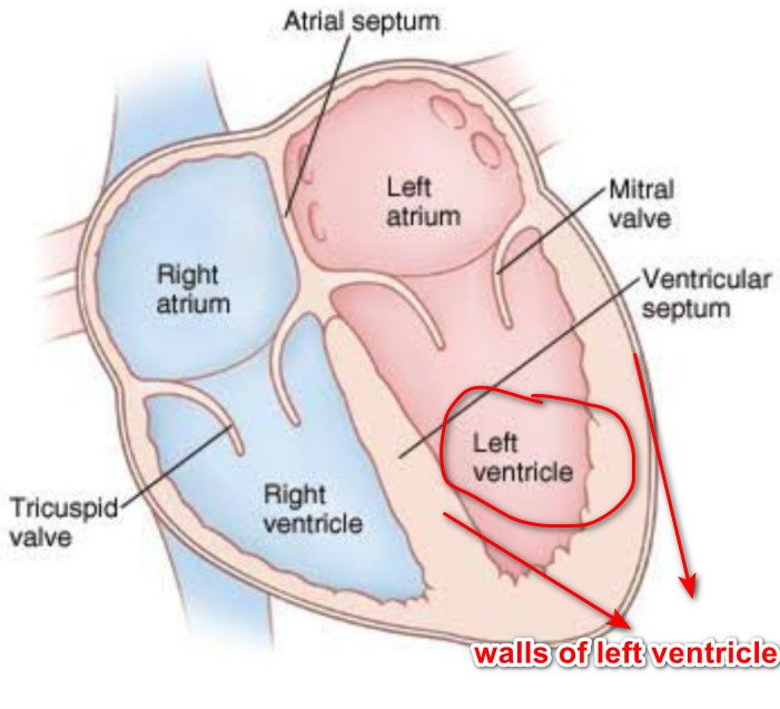
We can see here that left ventricle is on bottom of left side of the heart. RWMA term is usually applied to function of this chamber. Left
ventricle is shortly called as LV. In 2d echo, walls of LV are labelled as anterior, lateral, inferior and septal depending on the location of the wall. In RWMA, a particular wall among these is not contracting in the way it should be.

If blood supply to a part of heart is compromised severely, that part will not contract properly. it may show decreased contraction or no
contraction on 2d echo. This is labelled as regional wall motion abnormalities (RWMAs). Within seconds of cessation of blood flow to a part of heart due to blockage in the artery supplying that area, rwma develops. These changes occur prior to the onset of ECG changes or the development of chest pain. RWMAs were present in 93 percent of heart attack patients.
Causes for RWMA
- Heart attack
- Myocarditis
- Stress cardiomyopathy
But main application of rwma in clinical practice is for heart attack. But it may be impossible to distinguish RWMAs due to recent heart
attack from those due to a previous heart attack.
APPLICATION OF RWMA
RWMA can identify the location and extent of Heart attack or myocardial infarction
ANTERIOR RWMA— this is typical of occlusion of left anterior descending coronary artery circulation(LAD)
INFERIOR RWMA — It arises from occlusion of the right coronary artery (RCA)
LATERAL RWMA— Arises in circumflex artery occlusion (LCX)
In order to understand this, have a look at below picture

What happens to my heart if I have RWMA?
As that part of the heart is not contracting properly, it does not contribute much to the heart function. Function of your heart comes down. It means, heart becomes weak. Depending on the extent of RWMA and severity of RWMA, decline in heart function varies. RWMA suggest either ongoing myocardial infarction or previous myocardial infarction. ECH test and clinical history is useful to differentiate the both.
What tests will be advised if I have RWMA?
As coronary artery disease is the most common cause of RWMA, your doctor will advice coronary angiogram. Coronary angiogram looks for the blockages in coronary arteries. Your are likely to have blockages if you have RWMA.
Is there any treatment for RWMA?
If rwma is due to acute MI (myocardial infarction), you need immediate stent surgery to the heart, If that is not available, you will be given a drug into your vein to lyse the clot in the coronary artery. This is called thrombolysis therapy. After that , you need to continue certain tablets for rest of your life.
If rwma is due to previous myocardial infarction, you will be given tablets to improve the function of the heart, to prevent recurrent myocardial infarction and to improve life expectancy.
Conclusion is that RWMA represent decreased contractility of a wall of the heart. It occurs due to decreased blood supply to apart of the heart, it is detected by 2d echo. Rwma helps in diagnosing heart attack, it even helps in identifying extent of heart attack and artery involved in heart attack.